|
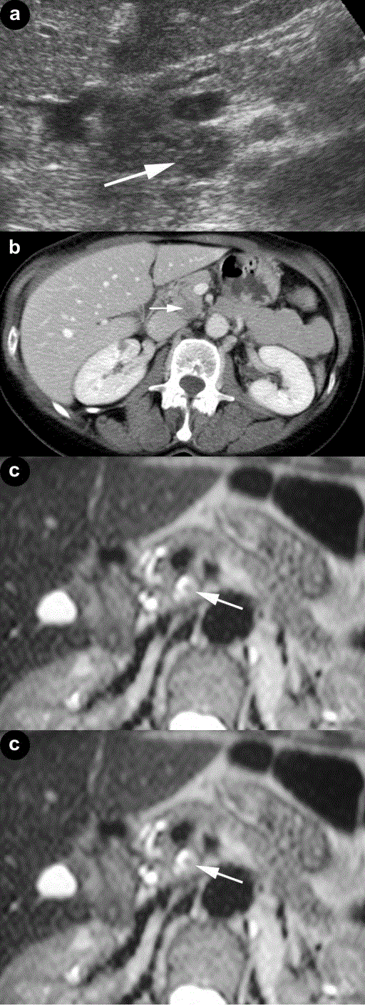
| Figure 1. Wrong diagnosis of IPMN at imaging. a. Ultrasound examination revealed the presence of a small microcystic lesion (arrow) of the pancreatic uncinate process with solid intralesional tissue and a slight dilation of the main pancreatic duct. b. At CT the lesion (arrow) was confirmed with a cystic appearance. MRI also confirmed the cyst which contained a small intralesional nodule (c. arrow) communicating with the main pancreatic duct (d. arrow). Moreover, a slight dilation of the main pancreatic duct was documented. |